Long QT Syndrome Exposed by Effort
AAM. Wilde, TA. Simmers.
Department of Cardiology. Academic Medical Centre.
University of Amsterdam. Amsterdam (The Netherlands)
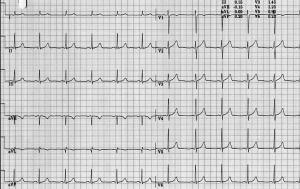
A 14-year old boy suddenly died while playing soccer. He was in the middle of a sprint when he suddenly succumbed. Resuscitation efforts were unsuccessful. His family assured that he had been without complaints before and that his family history was unremarkable. A two year older brother, however, remembered that he had also collapsed once while playing an exciting soccer match. This occurred at the age of 10, after which he experienced no further events. His brother’s death worried him (and his family) and he visited a cardiologist for medical advice Physical examination was unremarkable; his ECG is shown in Fig 1. The ECG shows sinus rhythm (70 bpm) with a normal QRS axis. PQ interval and QRS width are normal. Repolarization is completely normal and the QTc-interval is 384 msec, well within normal limits. Hence the ECG is completely normal. An echocardiogram was also normal.
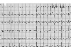
From the history of the patient and from his family history it became clear that both events (his collapse and the circumstances of his brother’s death) were triggered by exercise. An exercise test should therefore be part of the cardiological work-up. Figure 2 shows the ECG after 6 minutes of exercise. There is still sinus rhythm, 130 bpm, and conduction intervals remain normal. The QT-interval is now markedly prolonged and approaches 530 msec (QTc: 527 msec). This response should raise suspicion on a long QT syndrome, type 1 and in conjunction with the complaint(s) of the patient ß-blockade therapy is warranted. Molecular genetic screening indeed revealed a mutation in the KCNQ1 gene.
Editor's comments
The congenital long QT syndrome (LQTS) is a genetically transmitted disease. Mutations causing LQTS have been identified in 5 genes each encoding a cardiac ion channel and its regulatory subunit [1] . LQT1 is caused by mutations in the KCNQ1 gene encoding a potassium channel conducting the IKs potassium current. Detection of QTc prolongation in the basal ECG is the hallmark feature in the diagnosis of LQTS. However, studies that investigated QTc in genotyped LQTS families reported LQT1 patients having disease-causing mutations despite a normal QTc [2-4]. Type 1 LQTS is characterized by QT-prolongation, in particular during exercise. The QT-interval fails to adapt to an increase in rate and therefore inappropriately prolongs during exercise [5,6]. In conjunction, events (dizziness, syncope and sudden death) are typically triggered by adrenergic stimuli like exercise, diving and swimming [7], the age of onset of complaints is usually around 5 years. The present case exemplifies a LQT1 patient in whom QT prolongation only may be detected during an exercise test. In these cases, a careful family history should be taken and molecular genetic screening is mandatory. In symptomatic patients with LQT1, treatment of choice is a ß-blocker titrated up to the highest possible tolerated dose [8,9]. Asymptomatic young patients should receive prophylactic treatment but asymptomatic individuals over 20 years of age with a QTc interval < 500 msec seem to be at low risk (see also case III-6).
References
<biblio>
- Splawski pmid=10973849
- Vincent pmid=1508244
- Zareba pmid=9753711
- Moretti Moretti P; Calcaterra G, Napolitano C, et al. High prevalence of concealed long QT syndrome among carriers of KVLQT1 defects. Circulation 2001; 201 (Suppl): II-584 (abstract).
- Schwartz Schwartz PJ, Priori SG, Spazzolini C, et al. Genotype-phenotype correlation in the long-QT syndrome. Gene-specific triggers for life-treating arrhythmias. Circulation 2001; 103: 89-95.
- Villain pmid=15321698
- Priori pmid=11482917
<biblio>

